Edvotek® Restriction Enzyme Mapping
£132.78
£110.65
Explore restriction enzymes, plasmid DNA, and genetic mapping
Pack Size:
1
In stock
 Chilled
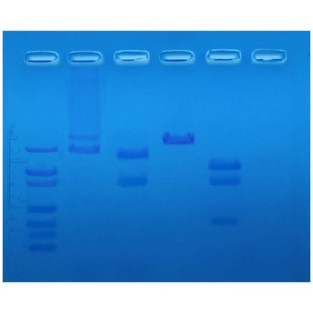
In this experiment, a plasmid DNA is cleaved with different combinations of restriction enzymes. By determining the fragment size and using agarose gel electrophoresis, the relative positions of the restriction sites can be mapped.
• Explore restriction enzymes, plasmid DNA, and genetic mapping
• Perform four DNA digestions and analyze the results using gel electrophoresis
• Create a standard curve, calculate the lengths of DNA fragments, and construct an enzyme map of the original plasmid
• A restriction enzyme lab with a strong analysis component!
Separation is achieved through an agarose gel, via an electric current. So, the molecules of DNA are separated based on their size and electrical charge. (The same technique is used for RNA separation, and protein separation, although the gel is orientated differently). After the tank is filled with enough running buffer (specific electrolyte solution) to cover the gel, the DNA fragments are loaded into wells in the gel that are at the negative electrode end of the gel tank. Coloured markers and carriers are added to the samples to aid loading (the carrier keeps the sample in the well) and visualization. The lid is added to the tank and current is allowed to flow from the negative (black) terminal to the positive (red) one. (Remember: “run to red or you’re dead!”).
Group Size: 6 sets of restriction digestions
Time Required: Complete in 1 hour 20 minutes to 1 hour 45 minutes
Kit Includes: Instructions, plasmid DNA for restriction digest, restriction enzyme reaction buffer, UltraPure water, restriction enzyme dilution buffer, EcoRI Dryzyme, BamHI Dryzyme, DNA standard marker, UltraSpec-Agarose™, electrophoresis buffer (50X), 10X gel loading solution, SYBR® Safe stain, FlashBlue™ stain, microcentrifuge tubes with attached caps
All You Need: DNA Electrophoresis, Micropipettes: 5-50 µl, Water Bath, UV Transilluminator (for InstaStain™ Ethidium Bromide), White Light Box (for FlashBlue™), & Microwave or Hot plate.
Storage: Some components require freezer storage upon receipt
| Pack Size | 1 |
|---|---|
| Gross Weight | 0.6 |
Write Your Own Review


